ABCDE assessment of an acutely unwell patient

James Hobson   Published 13th February 2023

On this page


The information on this page was accurate at the time of publication. Always consult current national and local guidelines. Content is for training purposes only.

The A-E Assessment

You’re an FY1 on shift in the Emergency Department and one of your patients has suddenly become very breathless. What do you do? Panic… Run to find your consultant… Try and find your second-year respiratory lecture notes…

Or, assess and manage them in a structured approach to acutely unwell patients?

An A-E, or ABCDE, assessment is a method of assessing and stabilising a patient in a stepwise approach - in order of priority of the things that will cause the most harm. You can perform an A-E assessment on any unwell or deteriorating patient to help you ascertain what’s going on.

When performing an A-E assessment:

  • Treat any abnormalities you find as you go
  • Reassess the patient regularly to see if your treatments have worked
  • Remember the A-E is designed to stabilise the patient before more definitive treatment of the underlying problem can be provided

Airway

“Hello, are you OK?”

Is the patient talking to you? If they’re responding to you, you know their airway is patent. Ensure to speak loudly into each ear in case the patient is deaf on one side. If you get no response, give a painful stimulus using trapezius squeeze or sternal rub to see if they respond to pain.

Always call for help urgently in an unresponsive patient.


Causes of airway compromise

  • Depressed level of consciousness - such as in opioid overdose, head injury and stroke
  • Fluid in the airway
    • Blood from epistaxis, haematemesis or trauma
    • Vomit/secretions in the airway from alcohol intoxication, head trauma or dysphagia
  • Inhaled foreign body
  • Damage to the airway from trauma to the face or throat
  • Swelling of the epiglottis from epiglottitis
  • Anaphylaxis – causes swelling of the upper airway (angioedema)
  • Other soft tissue swelling of the airway – such as infection (e.g. quinsy), venous outflow obstruction (e.g. front of neck haematoma)
  • Laryngospasm in asthma, gastro-oesophageal reflux disease (GORD) or from intubation
  • Local mass effect – such as tumours and lymphadenopathy (e.g. lymphoma)
  • Blocked tracheostomy


Look

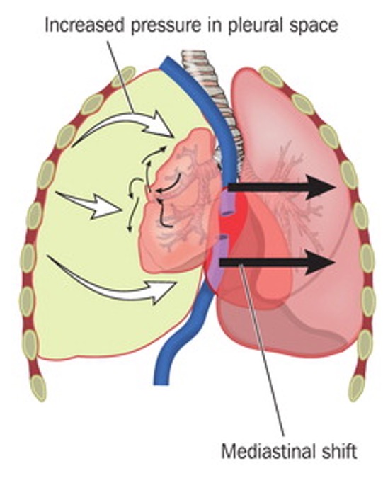
Indicative of an airway obstruction.

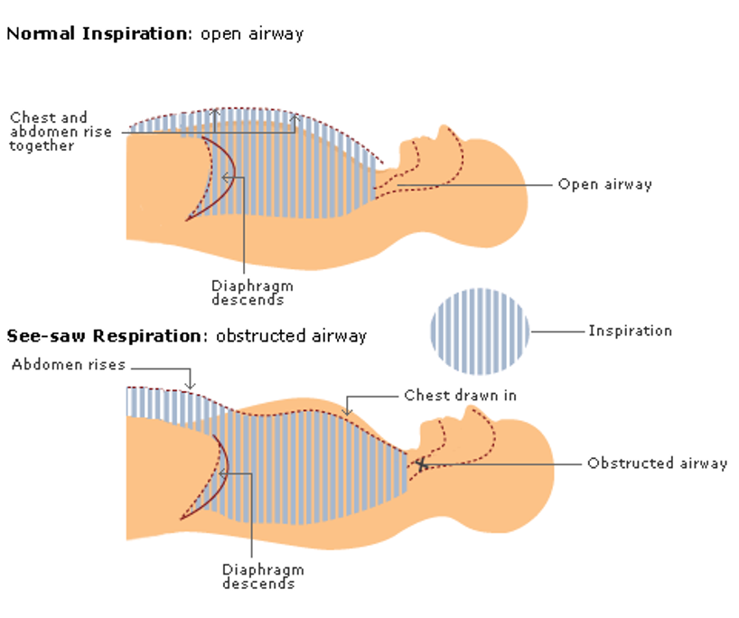
In inspiration, the diaphragm descends reducing intrathoracic pressure. During normal breathing with a patent airway this causes the external air (at a relatively high pressure) to be drawn into the lungs. The chest wall expands as the lungs fill with air

When the airway is obstructed, the diaphragm still descends, and the intrathoracic pressure is reduced. But this time no air can be brought in through the airway. Instead, the reduced pressure sucks the chest wall inwards. The lowering of the diaphragm gives the impression of the abdomen distending.

This is see-saw breathing - a paradoxical movement where the chest wall gets sucked inwards during inspiration.

Graphic depicting normal and see-saw respiration

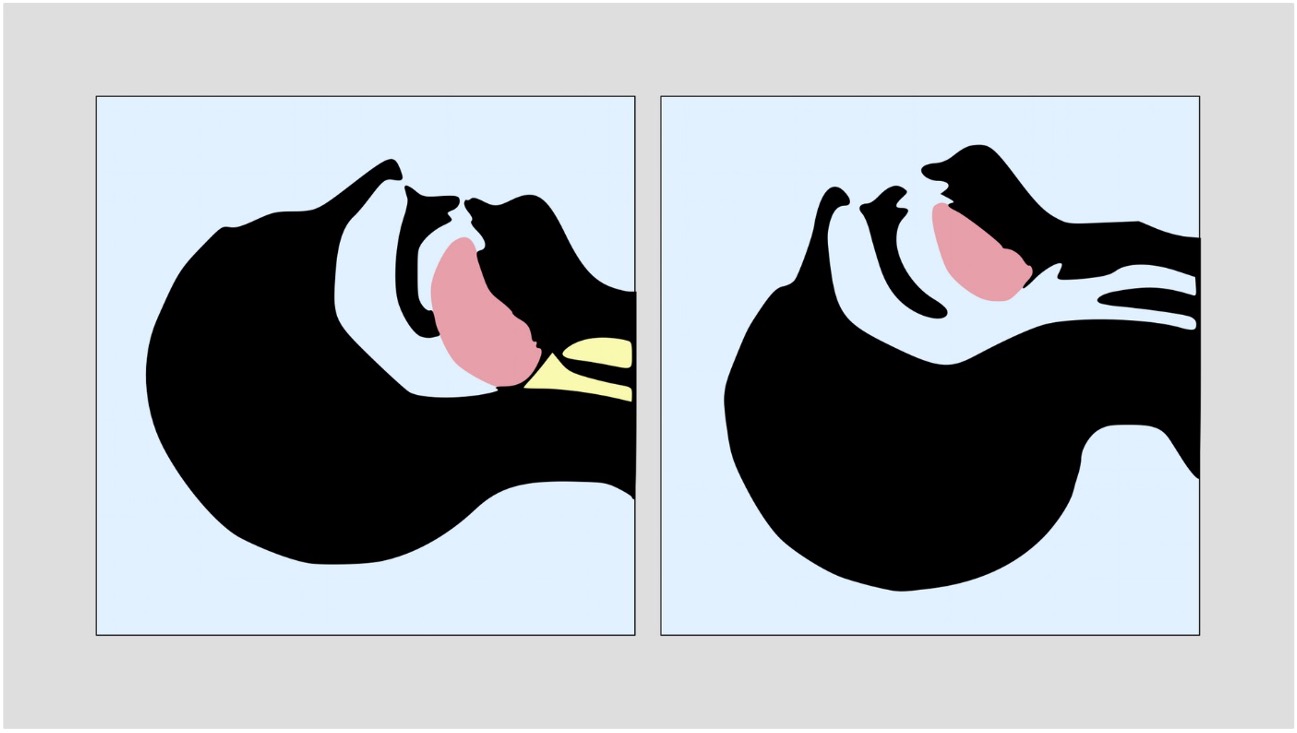
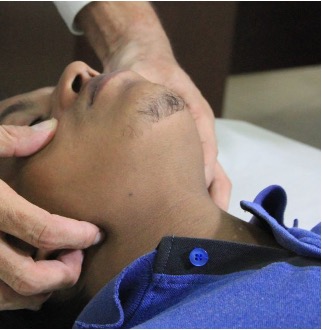
Check in the patients mouth for a visible obstruction such as a foreign body, vomit or secretions.


Feel

Place your cheek over the patients mouth

  • Lean over the patients mouth, looking down towards their feet. You can look for chest rise/fall, feel for breath on your cheek and listen for breathing
  • You should check for a maximum of 10 seconds. If the patient is not breathing, call for help and commence CPR

Listen

Absent breath sounds indicate a complete obstruction.

Additional breath sounds indicate a partial obstruction

A noisy, high-pitched, sound due to obstructed airflow in the larynx/trachea.

A low-pitched sound due to obstruction in the nasopharynx or oropharynx, sounds like nasal congestion or snoring.

High-pitched ‘musical’ sound when bronchial airways are narrowed – can be inspiratory, expiratory or biphasic. Wheeze can be heard across the lungs (e.g. asthma) or in a localised area (e.g. foreign body).

Secretions or other fluid (e.g. vomit or blood).


Interventions

Get expert help quickly!

Airways always need the most skilled practitioner available – usually an anaesthetist or an ITU/ED specialist - to maximise the chance of first-pass success for intubation. Excessive laryngeal manipulation from repeated attempts at intubation will lead to airway oedema and inflammation, making subsequent attempts more difficult.

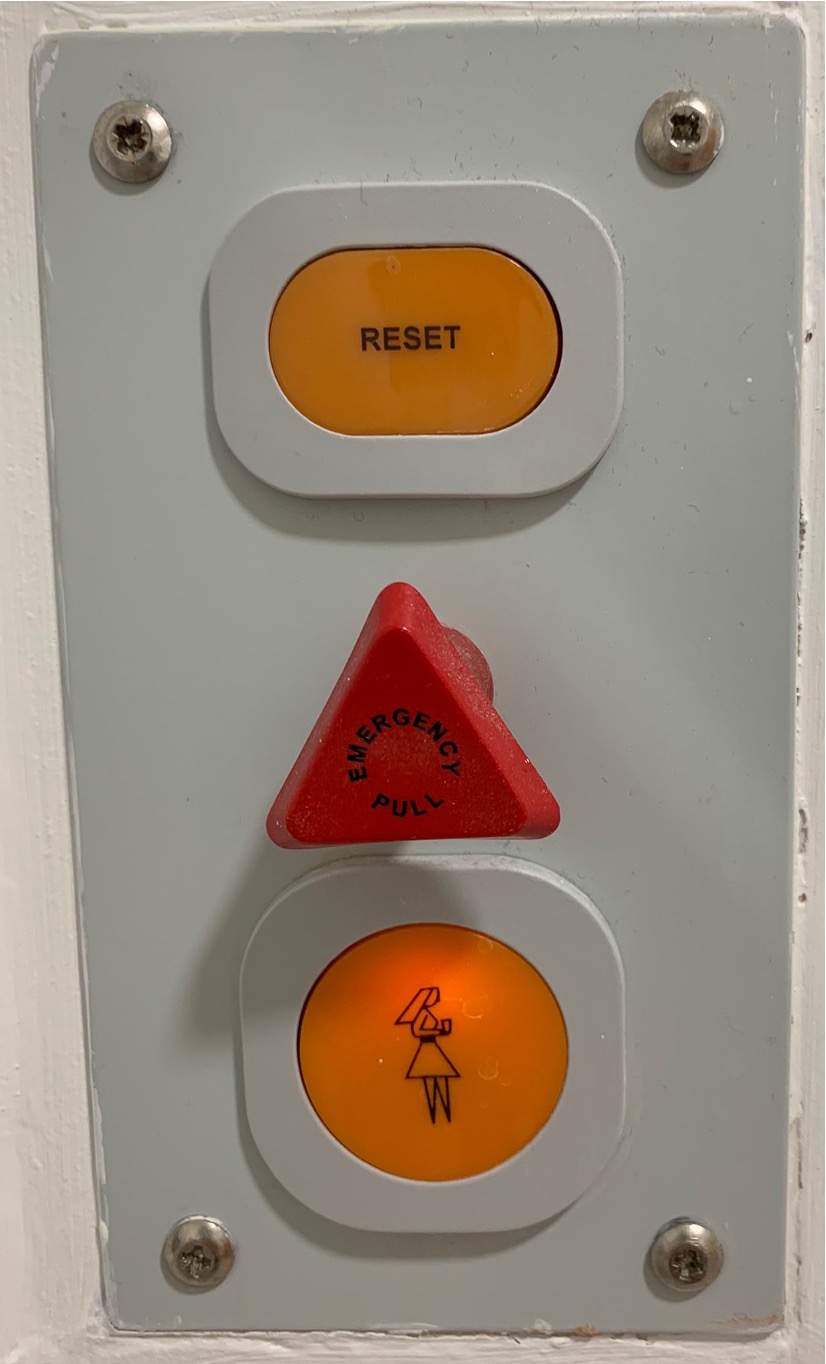
What will you do if there’s nobody around you? You should shout for help or pull the emergency alarm. Don’t be afraid to do this if you need to, nobody will judge you for getting help if you’re worried!

Hospital emergency alarm
Airways are like penises - the more you fool around with them, the harder they get

While you're waiting for help...

  • Perform manoeuvres to keep the airway patent - head-tilt chin-lift or jaw thrust
  • Remove any foreign bodies with magill forceps – no fingers in mouths!
  • Suction any vomit, blood or secretions as far as you can see
  • Ventilate the patient with a bag-valve mask
  • Insert an oropharyngeal (Guedel) or nasopharyngeal airway - help to maintain airway patency and can be used in conjunction with a bag-valve mask
  • Insert a supraglottic airway (i-Gel or laryngeal mask) if trained to do so
  • Have someone prepare the equipment for intubation and surgical airway

When help arrives...

  • They will be able to intubate the patient with an endotracheal tube - this is a definitive airway
  • Surgical airway (cricothyroidotomy or tracheostomy) can be performed in a situation where the patient cannot be intubated or ventilated (e.g. to bypass an obstruction)

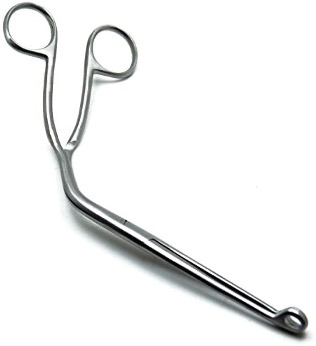
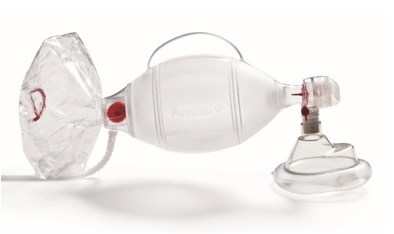
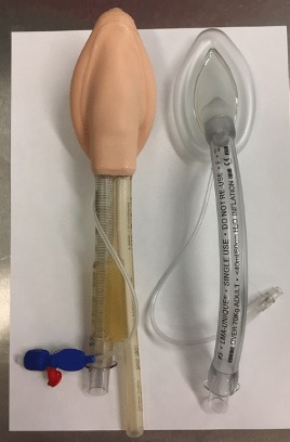
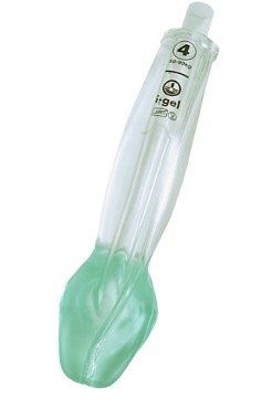
The following are techniques only to maintain airway patency:

  • Head-tilt chin-lift / jaw thrust
  • Removing foreign bodies / suction
  • Bag-valve-mask (BVM)
  • Oropharyngeal airway (OPA)
  • Nasopharyngeal airway (NPA)
  • Supraglottic airways – i-Gel or LMA

Whereas these also protect the airway:

  • Intubation with ETT
  • Surgical airway – cricothyroidotomy / tracheostomy

Protective measures maintain patency, but are also a completely closed circuit, so prevent secretions/vomit from entering the airway or air from inflating the stomach. They are used for definitive airway protection.


Head-tilt chin-lift
Head-tilt chin-lift
Jaw thrust
Jaw thrust
Nasopharyngeal Airway
Oropharyngeal Airway
Suction
Suction
Magill Forceps
Magill Forceps
Bag Valve Mask
Bag Valve Mask
Laryngeal Mask Airway
i-Gel
i-Gel
Endotracheal Intubation

Breathing

Causes of respiratory difficulty

Group causes based on:

  • Respiratory drive (the stimulation to breathe)
    • CNS Depression - loss of respiratory stimulation - e.g. opiate overdose, stroke
  • Respiratory effort (the ability to draw in breath by using the diaphragm and chest wall to manipulate intra-thoracic pressure)
    • Damage anywhere along the motor pathway supplying the muscles of respiration - C-spine, nerve roots, motor fibres (e.g. multiple sclerosis / Guillain-Barré Syndrome), neuromuscular junction (e.g. myasthenia gravis)
    • Restriction to ventilation - e.g. chest wall abnormality, pneumothorax, pleural effusion
    • Reduced respiration due to pain from rib fractures
  • Primary lung disorder (direct issue with the lung tissue)
    • Infection
    • COPD
    • Asthma
    • Pulmonary embolism
    • Pulmonary oedema

If breathing is insufficient to oxygenate the blood, the patient can quickly go into cardiac arrest.


Look

  • Shortness of breath (dyspnoea)
  • Cough
  • Signs of respiratory distress, such as:

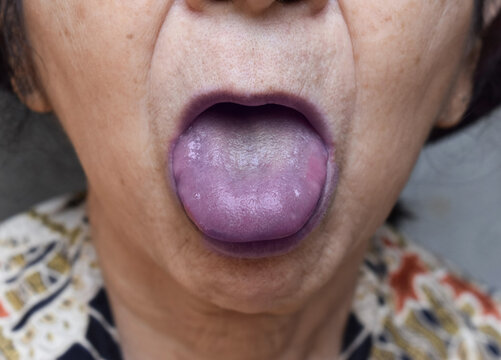
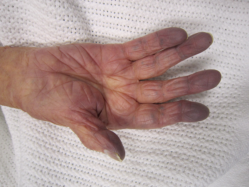
Cyanosis is a blue-purple discolouration caused by a lack of oxygen to the affected tissue. This could be due to poor oxygen uptake in the lungs, poor transport of oxygen bound to haemoglobin, or poor offloading of oxygen to tissues.

Commonly this affects the skin of the peripheries (e.g. fingers and toes) in peripheral cyanosis, or mucous membranes (e.g. lips and tongue) in central cyanosis.

  • Peripheral cyanosis is a result of reduced blood flow to the area (e.g. vasoconstriction due to cold exposure, peripheral vascular disease)
  • Central cyanosis is a result of diminished arterial oxygen (e.g. asthma, COPD, carbon monoxide poisoning, anaemia)

Central Cyanosis

Blue discolouration of the tongue and lips seen in central cyanosis

Peripheral Cyanosis

Blue discolouration of the fingertips seen in peripheral cyanosis

Normal inspiration is performed by the diaphragm and external intercostal muscles. Expiration is a passive process where the lungs recoil.

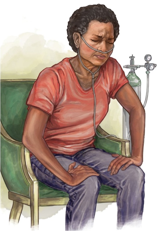
Using accessory muscles to aid breathing indicates increased work of breathing. Accessory muscles of respiration include the sternocleidomastoid, scalene, pectoralis major and trapezius muscles – these assist in inspiration. Abdominal muscles such as rectus abdominis and the obliques can assist with active expiration.

Sitting in the tripod position makes the use of accessory muscles easier, to help with increased work of breathing.

The image also shows pursed-lip breathing, which help to make breathing slower and more controlled.

Woman sat in the tripod position
Tripod positioning and pursed lip breathing

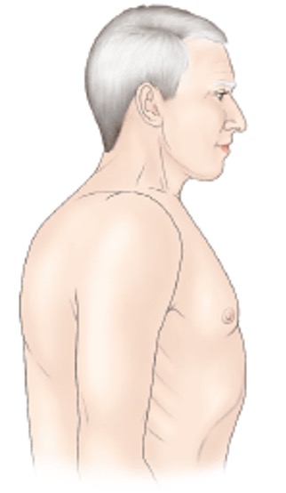
Classical finding in COPD. The lungs are unable to exhale fully due to reduction in elastic recoil and are therefore chronically overfilled with air.

Barrel chest
Barrel Chest


Feel

  • Tracheal deviation (e.g. in tension pneumothorax)
  • Chest expansion - is it equal?
  • Percussion note - dull or resonant?

Listen

  • Reduced breath sounds
  • Abnormal breath sounds - e.g. wheeze, stridor

Investigations

Respiratory investigations should focus on assessing lung ventilation and tissue perfusion.

For exams

When asked about investigations you would perform it is best practice to give them in order of invasiveness/complexity - i.e. start with things you can do at the bedside. If you jump straight to imaging you'll miss out the basics!

Think the five S's... bedSide tests (e.g. basic observations, ECG), Swabs (wounds, throat etc), Samples (bloods, urine, stool etc), Scans (radiological imaging), Surgery (biopsies, excisions and exploratory surgery)

  • Bedside tests
    • SpO2 and respiratory rate
    • Peak flow meter
  • Bloods/swabs
    • Arterial/venous blood gas
    • Sputum sample
    • Throat swabs
  • Imaging
    • Chest X-ray
    • Chest CT ± IV contrast

Interventions

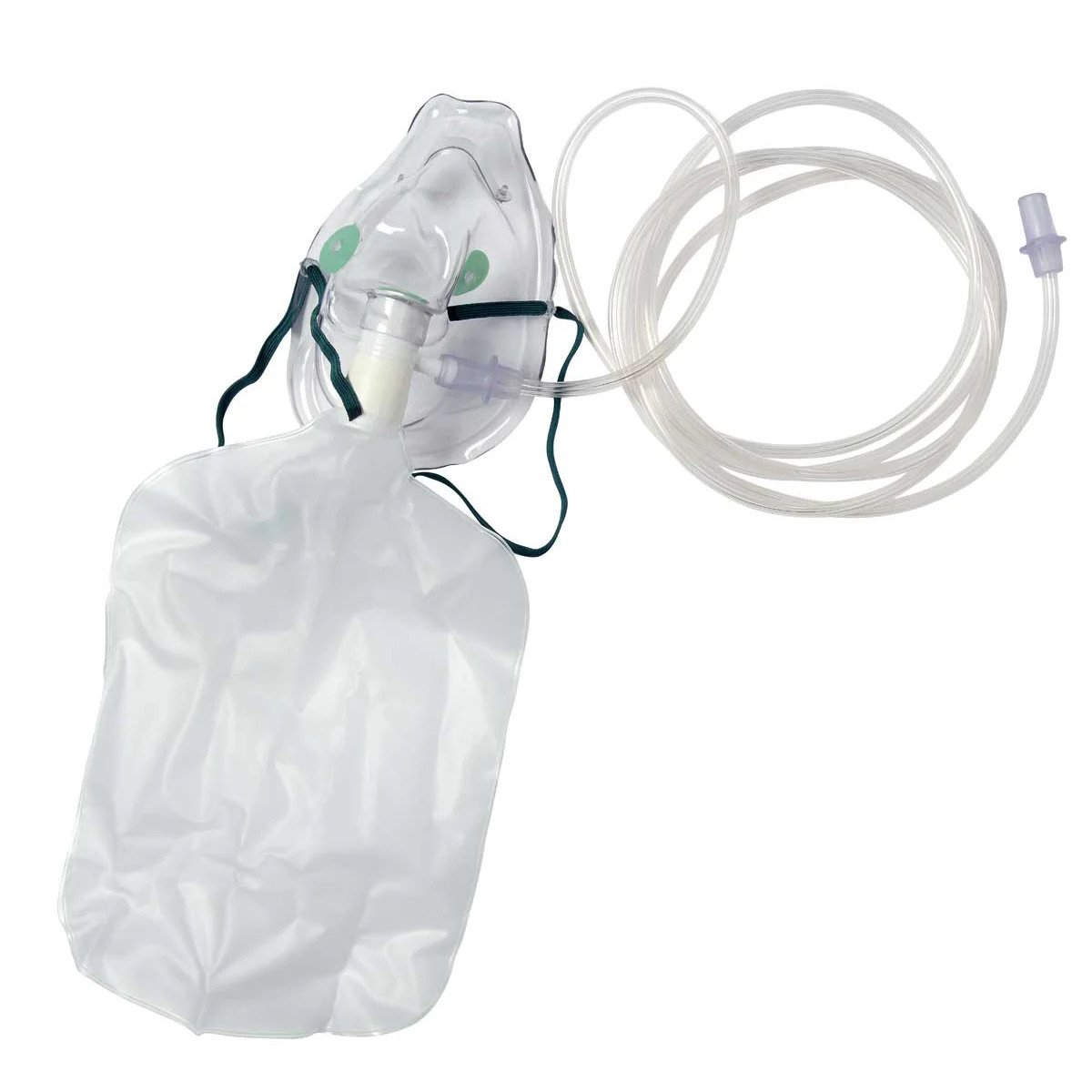
If the patient is hypoxic, provide supplemental oxygen 15L/min via a non-rebreathe mask and titrate according to response.

Patients with COPD who are unwell and hypoxic can still be started on 15L/min. Due to the risk of hypercapnia it is advised to use a 24-28% venturi mask and aim for O2 saturations 88-92%. ABGs allow monitoring of CO2 levels to assess CO2 retention.

Other treatments should be provided depending on the suspected cause, such as:

  • Tension pneumothorax - urgent needle decompression followed by cheat tube insertion
  • Asthma - nebulised SABA (e.g. Salbutamol) with SAMA (e.g. Ipratropium Bromide)
  • Anaphylaxis - IM adrenaline, steroids and antihistamines
  • Infection - consider sepsis six screen, start antibiotics
Non-Rebreathe Mask
Non-Rebreathe Mask

Circulation


Causes of circulatory collapse


Hypovolaemia

Reduced blood volume.

  • Dehydration - e.g. diarrhoea, vomiting, burns, DKA
  • Haemorrhage - loss of blood volume
  • Third-spacing - fluid leaves the intravascular space due to increased vascular permeability (e.g. due to release of inflammatory cytokines)
Patient with a bleeding wound on the arm being bandaged

Fluid distribution

Blood volume remains normal, but fluid is distributed to the wrong places.

  • Sepsis - inflammatory cytokines cause systemic vasodilation
  • Anaphylaxis
  • Spinal cord injury - blood vessels normally have a degree of sympathetic tone constricting them to ensure blood is directed to the correct places. If the sympathetic tone is lost due to neurological injury there will be mass vasodilation
Just ask could it be sepsis graphic

Cardiac pathology

Occurs in any condition where the heart cannot maintain adequate cardiac output.

  • Acute coronary syndromes - STEMI, NSTEMI, unstable angina
  • Aortic dissection
  • Arrhythmias
  • Cardiomyopathy
  • Myocarditis
Graphic of atherosclerotic plaque in a coronary artery

Obstruction

Obstruction of the heart's ability to pump or of the outflow of blood through the great vessels.

  • Pulmonary embolism
  • Tension pneumothorax
  • Cardiac tamponade
  • Coarctation of the aorta
Graphic depicting mediastinal shift in tension pneumothorax


Look

  • Pallor / cyanosis - sign of poor perfusion
  • Peripheral or pulmonary oedema - sign of circulatory overload
  • Sweating - sign of a sympathetic response (e.g. when compensating for hypotension)
  • Cold/clammy skin - sign of poor perfusion / peripheral vasoconstriciton
  • Mottled skin (livedo reticularis)
  • Blood loss


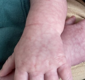
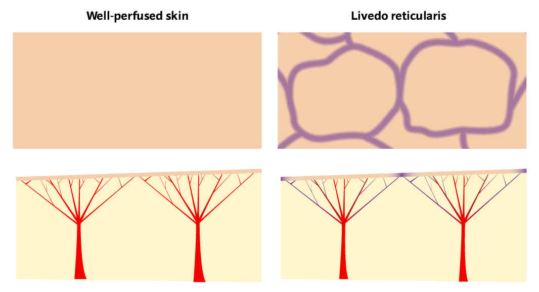
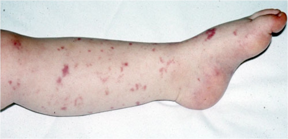
Mottling of the skin (livedo reticularis) is a classic skin finding in shock or other states of poor perfusion.

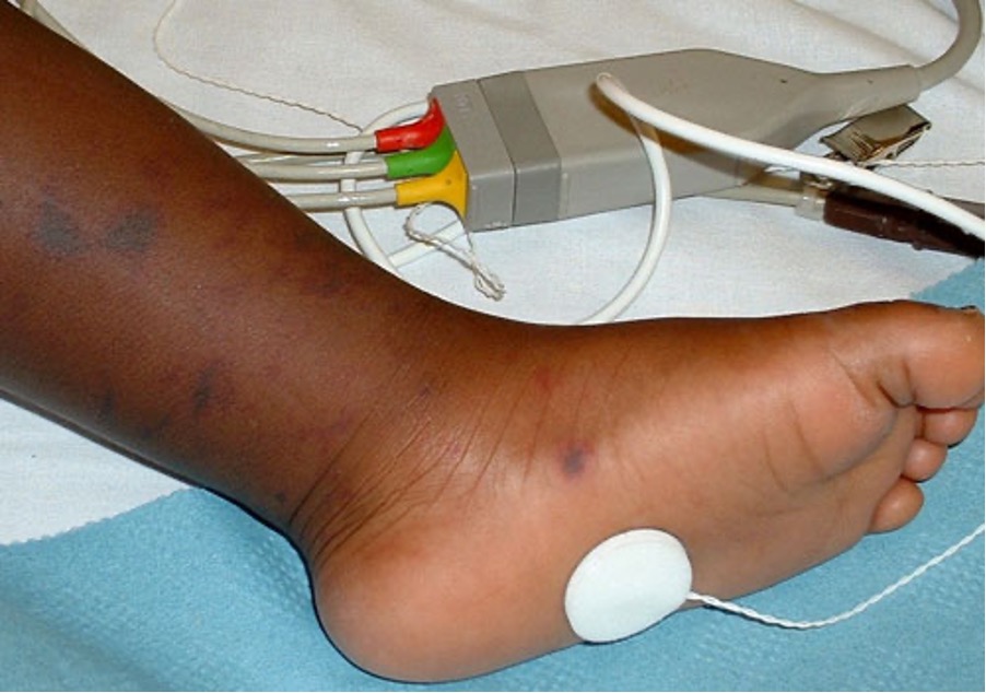
Each capillary bed is like a tree, the arteriole is the trunk with all the capillaries reaching out up to the skin. There are lots of these next to each other, each supplying a small area of the skin.

When there is poor tissue perfusion, the capillary beds furthest from the arteriole will be the first to be affected by the low oxygen saturation. The periphery of each capillary bed receives less oxygen and the skin it supplies begins to cyanose. But the centre of each arterial "tree" is still getting enough oxygen.

So looking from the surface you'll see a ring of cyanosis, with normal skin in the centre. With all these "trees" next to each other the pattern looks like a net of cyanosis (reticular pattern).

livedo reticularis
Livedo Reticularis
livedo reticularis pathogenesis
Pathogenesis of Livedo Reticularis

Feel

  • Compare temperature of the peripheries bilaterally - do they feel cold?
  • Assess capillary refill time (CRT) - squeeze the fingertip for five seconds, on releasing the colour should flush back within two seconds
  • Pulse - it is important to feel the pulse (as well as getting a rate measurement on monitoring) to assess the rate, rhythm (regular or irregular) and character (normal, weak, thready, bounding etc)

Listen

  • Heart sounds - listen in each valve area. It is recommended to feel the pulse at the same time to easily identify S1 and S2, and categorise any added sounds as systolic or diastolic


Investigations

  • Bedside tests
    • Heart rate
    • Blood pressure
    • Core temperature
    • Capillary refill time
    • Assess jugular venous pressure (JVP)
    • Assess fluid balance
    • 12-lead ECG
  • Blood tests - insert at least one wide-bore cannula and take bloods, such as:
    • Full blood count - are they anaemic? Can you classify the anaemia? Signs of infection? Is it likely bacterial or viral?
    • Urea & electrolytes - baseline renal function for medications and fluids, identify and monitor electrolyte disturbances
    • Liver function tests - baseline is important for dosing many medications, especially antibiotics
    • C-reactive protein - non-specific inflammatory marker, raised in infections
    • Lactate - lactate is the end product of anaerobic glycolysis. It can be a good measure of end-organ damage in hypoxic states (e.g. sepsis or shock), poor perfusion (e.g. dehydration) or poor lactate clearance (e.g. liver damage)
    • Blood cultures - essential in ?sepsis - what organisms can be grown and what treatments are they sensitive to?
    • Troponin T/I - high sensitivity biomarker of cardiac injury used primarily in acute coronary syndromes
    • NTpro-BNP - biomarker for heart failure, secreted by an over-stretched ventricle and has a diuretic effect
    • Coagulation studies - includes PT/APTT/fibrinogen, can add on INR calculation
    • D-dimer - fibrin degradation product raised in any coagulative/fibrinolytic process (e.g. PE, DVT, DIC) - high sensitivty but low specificity (also raised in malignancy, infection, pregnancy, after surgery etc)
  • Imaging
    • Echocardiogram - echo can usually be performed with bedside ultrasound in the Emergency Department
    • Vascular ultrasound - USS with doppler for assessing vessel continuity and patency (e.g. to rule out abdominal aortic aneurysm or DVT)


Gauge and flow rate have no real correlation, gauge is an arbitrary number.

The difference between 18G and 16G doesn’t sound particularly significant. But 16G actually has twice the flow rate of 18G. This makes a huge difference for rapid fluid replacement and resuscitation.


Gauge Colour Flow rate
14G Orange 240 ml/min
16G Grey 180 ml/min
18G Green 90 ml/min
20G Pink 60 ml/min

Day-to-day on the wards you will see most patients with a pink cannula (20G) for giving normal slow IV infusions. You may see blue cannulas (22G) for older adults with fragile veins. Grey or orange cannulas are considered "wide-bore" and should always be used for resuscitation, rapid fluid replacement, blood transfusion, surgery and trauma.


Interventions

Fluid resuscitation

If the patient has low blood pressure (or signs of compensated shock), or fluids form part of a treatment protocol (e.g. hyperglycaemia) then start fluid resuscitation through the cannula you inserted.

Don’t give fluid unnecessarily due to the risk of circulatory overload

  • Give normal saline or Hartmann’s solution – 500ml over 15 mins
  • If at risk of fluid overload (e.g. known congestive heart failure) - start with a 250ml bolus
  • Fluid resuscitate up to a maximum of 2000ml – if you still need to volume replace beyond this you’re likely to need blood products
    • Think about giving fluid replacement like topping up orange squash with water... if you dilute it too much you won't be able to taste it, you'll have to add more squash to concentrate it again
    • The same goes for blood, give too much fluid and you'll be diluting the red blood cells, from then on you'll need to top up with packed red cells instead. Fluid replacement is essential for maintaining blood volume, but don't forget saline can't carry oxygen!

Blood transfusion

Blood products should always be given in haemorrhage, it is best practice to replace 'like-for-like' where possible.

In some settings, especially trauma surgery, autologous blood transfusion can be performed. As blood is suctioned from the surgical field, a cell salvage machine can extract the red blood cells and recycle them to be transfused back into the patient.

Vasopressors and inotropes

These are drugs which cause peripheral vasoconstriction (vasopressors) and increase the force of contraction of the heart (inotropes). They may act solely as a vasopressor or inotrope, or perform a mix of both actions.

Examples include: adrenaline, noradrenaline, dopamine, phenylephrine, ephidrine.

They should only be initiated after consultation with a senior and under supervision.

Haemorrhage

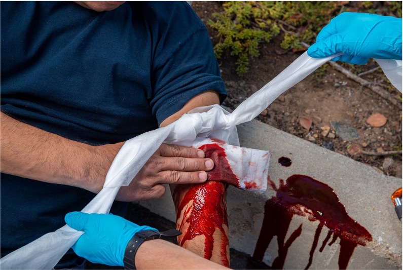
Take haemostatic measures.

  • Apply direct pressure, pack the wound and apply a tourniquet if required
  • Give tranexamic acid (TXA) - an antifibrinolytic drug to prevent clot breakdown
  • Give local drug agents to area (e.g. adrenaline-soaked gauze - causes local vasoconstriction)
  • Cauterisation or ligation of blood vessels
  • Reverse anticoagulants
  • Activate the massive haemorrhage protocol if required

Treat based on the likely causes:

  • Acute coronary syndrome - Morphine/Diamorphine, Oxygen, Nitroglycerine, Aspirin and antiplatelet agent (e.g. Prasugrel or Clopidogrel)
  • Cardiac tamponade - pericardiocentesis
  • Tension pneumothorax - needle decompression and chest tube
  • Anaphylaxis - IM adrenaline
  • Adrenal crisis - IV hydrocortisone
  • Hypertensive crisis - IV antihypertensives

The primary focus of circulatory issues is on shock (when blood pressure is low). Don't forget it is also possible to get a hypertensive crisis with a high blood pressure, requiring antihypertensive treatment.

Disability


Causes of reduced neurological status

  • Hypovolaemia
  • Hypoxia
  • Hypercapnia (e.g. COPD)
  • Metabolic disturbance (e.g. hypoglycaemia, adrenal crisis, myxoedema coma from severe hypothyroidism)
  • Epilepsy
  • Raised intracranial pressure or other neurological insults (e.g. stroke, traumatic brain injury, cerebral contusion)
  • Infections (e.g. meningitis, encephalitis)
  • Drug overdose (e.g. opiates, benzodiazepines, barbiturates)/toxins (e.g. carbon monoxide)
  • Iatrogenic causes (e.g. administration of high-dose opiates to an opioid-naive patient)
  • Delirium (important to consider in older patients, secondary to underlying cause)
    • UTI
    • Electrolyte disturbance
    • Pneumonia
    • Polypharmacy
    • Constipation
    • Urinary retention
    • Pain
    • Change of environment/routine
Finger prick blood glucose monitor
Capillary blood glucose monitor


Assess neurological status

  • Calculate their AVPU/GCS score
    • AVPU - Are they alert? Do they respond to voice? Do they respond to pain? Are they unresponsive?
    • GCS - Glasgow Coma Scale - a more complex assessment of eye response, motor response and verbal response
  • DEFG - DON’T EVER FORGET GLUCOSE! (and ketones if glucose is elevated)
  • Pupillary response - are they equal and reactive to light?
  • Check the patient's temperature if not already done in circulation
  • Medications - check their drug chart! What medications are they prescribed vs what have they taken?
  • Toxins - alcohol and drugs
  • Pregnancy - could they be pregnant? Perform a urine pregnancy test
  • Neurological signs - e.g. hemiplegia, seizures, weakness, speech/vision/hearing loss, slurred speech
  • Signs of increased ICP – headache, confusion, vomiting, papilloedema, irregular breathing
  • Signs of head trauma – head wound, racoon eyes, CSF otorrhoea/rhinorrhoea, haemotympanum
    • Do they meet CT head criteria?
Glasgow Coma Scale
Glasgow Coma Scale (GCS)


Interventions

Can they maintain their airway?

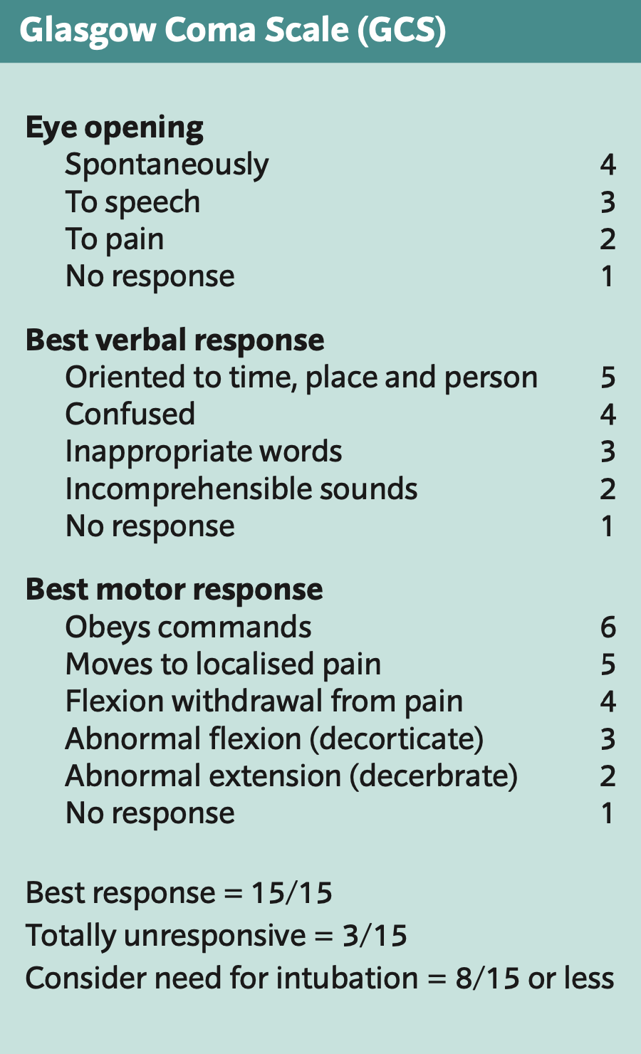
If the patient is unresponsive or only responsive to pain (GCS below 8), intubation is usually necessary.

  • Beware raised ICP, it makes airways risky!
  • Take measures to reduce ICP (e.g. raise head of bed, hyperosmolar therapy with hypertonic saline or IV mannitol, dexamethasone)
  • Excessive laryngeal manipulation leads to a sympathetic response which makes raised ICP worse, you always need the most experienced provider possible to get first-pass success
  • Induction agents (e.g. propofol) lower blood pressure, which reduces cerebral perfusion – use etomidate or ketamine to induce anaesthesia as these maintain blood pressure

Treat the causes:

  • Status epilepticus - generalised seizure for more than 5 mins, or >3 seizures in 1 hour with no recovery in between - benzodiazepines (e.g. IV lorazepam, IM midazolam, PR diazepam)
  • Hypoglycaemia - oral glucose or IV dextrose
  • Hyperglycaemia - assess for DKA/HHS, fluids, insulin, K+ replacement
  • Intoxication - is there a reversal agent/antidote? (e.g. naloxone for opiates, 100% oxygen for CO)
  • Haemorrhage – attempt to reduce ICP, manage symptoms and refer to neurosurgery

Exposure


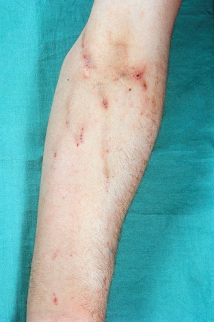
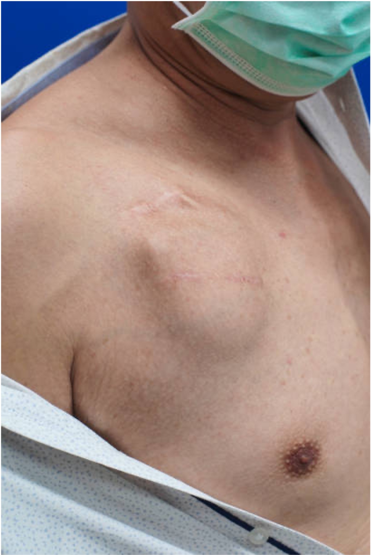
Perform a rapid whole-body inspection to check for other injuries or clinical signs – from top to bottom, front to back.

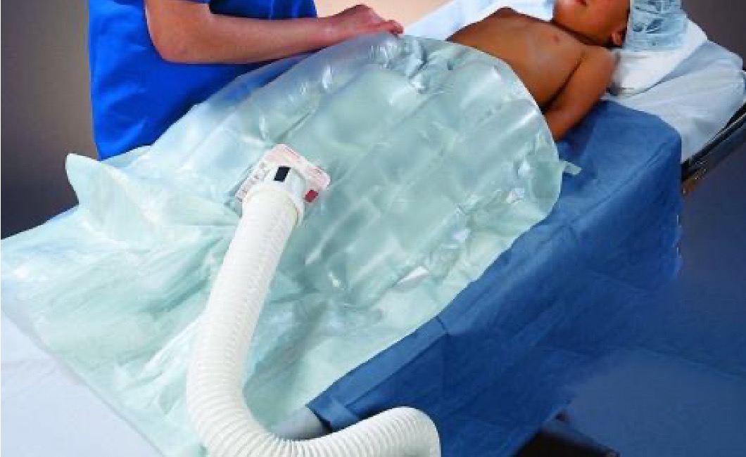
Expose the patient appropriately:

  • Make sure you check everywhere – regions often neglected include the scalp, the back, the orifices, the axillary, inguinal, and perineal regions, and body parts underneath dressings
  • Maintain the patient’s dignity and preserve body heat by only uncovering the areas you are examining in turn
  • Maintain C-spine precautions if relevant (e.g. by using the log roll manoeuvre)

Assess for injuries and clinical signs

  • Do they have any pain anywhere?
  • Inspect the skin for: wounds, rashes, bruises, swelling, erythema, tenderness, needle track marks, medication patches
  • Inspect any IV access in situ for: erythema, swelling, discharge, extravasation
  • Inspect the calves for signs of DVT or peipheral oedema
  • Active bleeding - estimate blood loss and rate of loss - then re-assess circulation for signs of shock

Purpuric rash
Purpuric/petechial rash of meningococcal sepsis on light skin
Purpuric rash
Purpuric/petechial rash of meningococcal sepsis on dark skin
Log roll
Log roll - for examining a patient with C-spine precautions
Needle track marks
Needle track marks due to intravenous drug use
Pacemaker
Pacemaker under the skin
Bair Hugger
Bair Hugger™ device for warming a patient

Interventions

If you've found any more clinical signs, check you're still treating for the most likely diagnosis. Focus on treating the unerlying condition (e.g. sepsis).

  • Remove wet/contaminated clothing and provide clean/dry clothes
  • Remove allergens/medications (e.g. insect stings, IV infusions)
  • Get swabs and cultures of any wounds
  • In hypothermia, start rewarming – initially with warm blankets or Bair Hugger™. You can also give warm IV fluids. In severe cases, thoracic/peritoneal cavity lavage, warm air ventilation and extracorporeal blood warming can be performed
  • Hyperthermia – surface cooling, cool IV fluids
  • DVT – Well’s score, D-dimer, lower-leg ultrasound, anticoagulation


Post-ABCDE Actions


Once you've finished your first pass of the A-E assessment you can focus more on what led to this deterioration event.

  • Keep re-assessing the patient and be prepared to escalate treatment if required
  • Take a focussed history from the patient – SAMPLE history
    • Symptoms/Signs
    • Allergies
    • Medications
    • Past medical history/pregnancy status
    • Last meal/drink/menstrual period/opened bowels and bladder
    • Events leading up to deterioration
  • Take a collateral history where possible (e.g. friends, family, carers, ward staff) to fill in any blanks
  • Review the patient’s notes, charts, labs, investigations, medications etc
  • Record what you’ve done and the patient’s response to treatment
  • Refer for further investigations/review from specialists and handover to the next team on shift using the SBAR approach