Simulation case | A 69 year old with chest pain

James Hobson   Published 10th February 2023

Handover from ED triage

John McKenna - 69yr old male attending with chest pain. He has a past medical history of type two diabetes, hypertension, and gastro-oesophageal reflux – for which he takes metformin 500mg TDS, amlodipine 5mg OD, omeprazole 20mg OD and Gavisgon liquid PRN after meals.

On further questioning:

  • The pain started ~1 hour ago in the centre of his chest. He tried taking some Gavisgon but it didn't help. He would now rate it 8/10.
  • John is feeling nauseous but hasn't vomited. He denies any palpitations, dyspnoea, cough, fever, fatigue or bladder/bowel changes.
  • No known drug allergies
  • He is an ex-smoker who stopped ~10 years ago. He has alcohol occasionally for special occasions.
  • His brother had a "clot on his lung" while in hospital last year, he's worried he might have the same thing.

HR 95, BP 142/91, RR 22, SpO2 91% OA, Temp 36.5C, Alert

Airway

Examination

John is talking. His airway is patent and self-maintaining.

Investigations

None required.

Interventions

None required.

Breathing

Examination

RR: 24 SpO2: 90%

Chest expansion – normal and equal

Percussion - normal resonance

Auscultation - good air entry bilaterally with no added sounds

Investigations

Students may ask for:

  • ABG
  • CXR

Interventions

Recognise hypoxia

  • Give oxygen – 15L/min via NRB – can titrate down based on patient response
  • Sitting the patient more upright may assist with ventilation

At this stage it may be appropriate to move the patient to ED Resus

Arterial Blood Gas for John McKenna


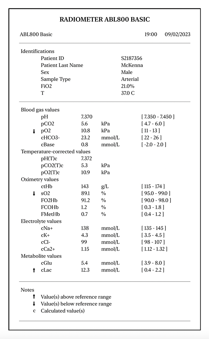
PaO2 is mildly reduced.

Lactate is elevated, as anaerobic glycolysis is taking place due to ischaemia.
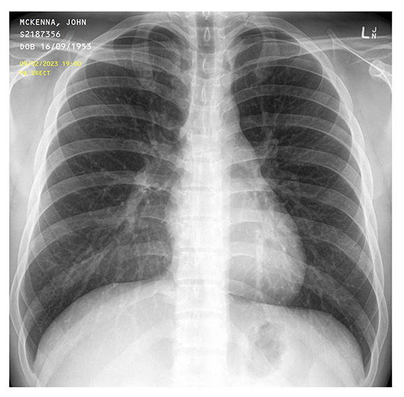
Chest X-Ray for John McKenna


Normal chest x-ray.

Circulation

Examination

HR 102 BP 140/90 T 36.5 C

John is pale and feels clammy

Pulse is fast and regular

No peripheral oedema

HS I + II + 0

If asked about urine output – last emptied bladder at home around 2 hours ago

Investigations

Insert at least one wide-bore cannula – 14G (Orange) or 16G (Grey)

Bloods – ask to justify:

  • FBC – anaemia, infection
  • U&Es – renal function and electrolyte levels
  • LFTs – liver function for medications
  • CRP – infection
  • Troponin – ?MI
  • Coags – assess for coagulopathy – as patient is likely to need PCI and long-term antiplatelet therapy

12-lead ECG

Interventions

Patient is not hypotensive so no fluids required for now.

Think ?STEMI:

  • Morphine (5-10mg IV)
  • Nitrates (GTN sublingual spray)
  • Aspirin (300mg PO loading dose)
  • Antiplatelet agent - Prasugrel (60mg PO) is preferred if undergoing PCI
  • May also consider anticoagulation with unfractionated heparin

Starting long-term prevention with beta-blockers, ACEi, statins etc is not indicated in A&E.

ECG for John McKenna


  • Rate ~82bpm
  • P waves precede QRS = sinus rhythm
  • Rhythm is regular
  • ST segment elevation is present in leads I, aVL, V1, V2, V3
  • Reciprocal ST segment depression is present in leads II, III and aVF
  • The ST elevation is in the anteroseptal and lateral leads (anterolateral STEMI), which represents a likely Left Anterior Descending Artery occlusion

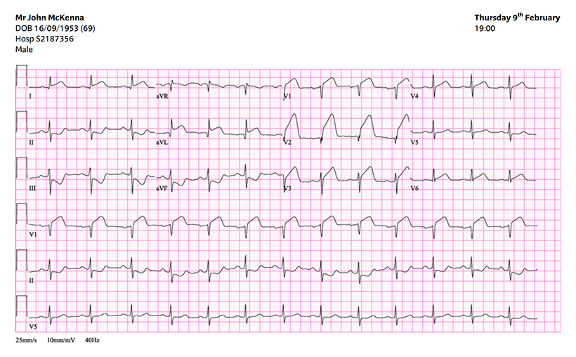
ECG criteria for STEMI

Persistent ST segment elevation in at least two contiguous leads of ≥1mm in all leads other than V2-V3.

In V2-V3 there must be ST segment elevation of:

  • ≥2.5 mm in men under 40
  • ≥2 mm in men over 40
  • ≥1.5 mm in women of any age

Blood results for John McKenna

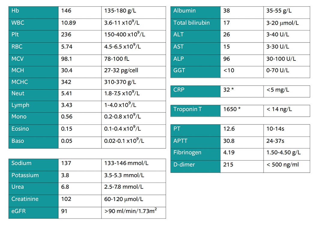
Troponin T is highly elevated - this is a sensitive biomarker of cardiac damage

CRP is elevated, this is an inflammatory marker and it is normal to be raised in STEMI

Disability

Examination

Alert, GCS 15/15

PEARL

Investigations

CBG – 5.2 mmol/L

Interventions

None required.

Exposure

Examination

Patient is pale and clammy

No skin findings

Abdomen SNT

Calves SNT

No peripheral oedema

Investigations

None required.

Interventions

None required.

Post-ABCDE Actions

Can you confirm a diagnosis?

What will you do now?

  1. Re-assess the patient regularly (Now SpO2 94% BP 128/80 HR 84 Pain 7/10)
  2. Verify diagnosis with senior in ED or discuss with CCU
  3. Call 999 and state “ambulance for time-critical transfer of STEMI patient from LRI ED Resus to GGH CCU”
  4. Call interventional cardiology red phone on 3222 stating ‘STEMI alert’ to handover patient
  5. Full history and relevant physical examination in the meantime
  6. Document in the patients notes

Handover using SBAR technique

  1. “Hello my name is X, I am calling from ED at Leicester Royal Infirmary. I have a patient, John McKenna, with ongoing anterolateral STEMI.
  2. The patient is a 69 year old male admitted with acute-onset constant severe chest tightness. He has a background of both hypertension and type 2 diabetes.
  3. On admission, he was alert, but hypoxic, hypertensive and tachycardic. I have given him supplementary oxygen, morphine for analgesia, GTN spray and loading doses of aspirin and prasugrel. He has one cannula in situ. Troponin T is raised at 1650 ng/L and ECG suggests anterolateral STEMI.
  4. Can you please arrange transfer for primary PCI for this patient.”